
Early Activated Microglia: The First Cells to Sense Alzheimer’s Disease
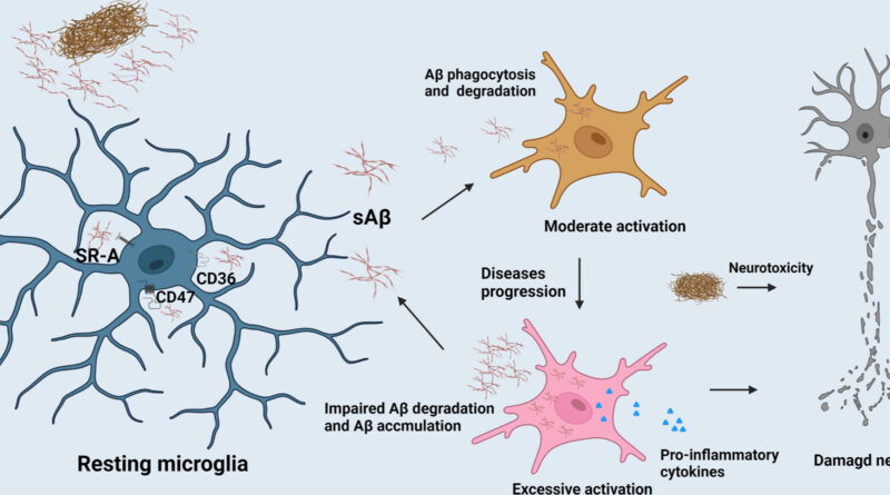
Early activated microglia are now recognized as the first cells to sense the earliest biological disruptions that precede Alzheimer’s disease. Long before neurons begin to fail, these microglial subpopulations detect subtle metabolic stress, early amyloid fragments, and synaptic instability.
In the vast landscape of the human brain, where billions of neurons fire in patterns that create memory, perception, and identity, scientists have long searched for the earliest signs of Alzheimer’s disease. For decades, the focus rested on neurons—their death, their loss of synapses, their accumulation of toxic proteins. But in the last few years, a quiet revolution has unfolded. Using single-cell RNA sequencing, spatial transcriptomics, and high-resolution imaging, researchers have discovered that the first cells to sense Alzheimer’s are not neurons at all. They are microglia—the brain’s immune sentinels, constantly patrolling the neural environment, responding to subtle changes long before symptoms appear.
Early Activated Microglia in the Earliest Stages of Alzheimer’s Disease
This discovery did not arrive suddenly. It emerged gradually, through a series of studies published between 2024 and 2026 in Nature, Nature Neuroscience, Cell, and Neuron. These studies, conducted by teams at the Broad Institute, MIT’s Picower Institute for Learning and Memory, UCSF’s Weill Institute for Neurosciences, Harvard Medical School, and the NIH BRAIN Initiative, converged on a single conclusion: microglia undergo distinct molecular transitions in the earliest phases of Alzheimer’s, forming a sub-population now known as Early-Activated Disease-Associated Microglia, or EADAM.
Unlike neurons, which begin to fail only after years of accumulating pathology, microglia respond almost immediately to the first signs of trouble. They detect subtle shifts in the extracellular environment—changes in lipid composition, early amyloid fragments, metabolic stress, and faint signals of synaptic dysfunction. Their response is not random. It is structured, coordinated, and deeply connected to the architecture of the disease.
The story begins with single-cell RNA sequencing, a technology that allows scientists to examine the gene expression of individual cells with extraordinary precision. When researchers applied this technique to brain tissue from early-stage Alzheimer’s patients and mouse models, they found that microglia were not uniform. Instead, they formed distinct clusters, each with its own molecular signature. Among these clusters, one stood out: a group of microglia expressing genes associated with interferon signaling, lipid metabolism, synaptic pruning, and early inflammatory responses. These cells appeared before widespread amyloid plaques, before tau tangles, and before neuronal death. They were the first responders.
Spatial transcriptomics added another layer of insight. By mapping gene expression directly onto brain tissue, scientists discovered that EADAM microglia cluster around vulnerable synapses, especially in the entorhinal cortex and hippocampal CA1 region—areas known to be the earliest affected in Alzheimer’s. They appear near synapses that are beginning to lose efficiency, near blood vessels showing early signs of breakdown, and near neurons experiencing subtle metabolic stress. Their presence forms a mosaic of early pathology, a cellular map of the disease’s hidden beginning.
What makes EADAM microglia so important is not just their early activation, but their function. Microglia are responsible for synaptic pruning—removing weak or unnecessary synapses to maintain neural efficiency. In healthy brains, this process is essential. But in Alzheimer’s, EADAM microglia appear to prune too aggressively, removing synapses that are still functional. This early synaptic loss may be one of the first cognitive disruptions in the disease, occurring long before memory loss becomes noticeable.
This idea resonates with zemeghub article Blood Biomarkers for Alzheimer’s: New Evidence May Detect the Disease in Its Earliest Stages — What Scientists Have Discovered. Several proteins associated with EADAM activation—especially those involved in interferon signaling and lipid metabolism—have already been detected in blood plasma years before symptoms appear. GFAP, a marker of astrocyte reactivity, is one of the strongest early biomarkers, but microglial markers are emerging as equally promising. The connection between microglial activation and blood biomarkers is becoming one of the most powerful avenues for early detection.
Why Early Activated Microglia Are Considered the First Responders in Alzheimer’s
The discovery of EADAM microglia also intersects with research on perception and reality construction. In your article How the Brain Constructs Reality — The Astonishing Science Behind Conscious Perception, you explored how the brain integrates sensory information into a coherent experience. Microglia play a subtle but essential role in this process. By regulating synaptic strength and pruning, they help maintain the fidelity of neural circuits. If they become overactive, the brain’s ability to construct stable perceptions may begin to falter. This could explain why individuals in the earliest phases of Alzheimer’s sometimes experience slight changes in attention, sensory integration, or cognitive flexibility long before memory loss becomes severe.
Stress adds another dimension. In zemeghub article The Brain Under Extreme Stress — What Scientists Discovered in Real Crisis Situations, you described how stress reshapes neural circuits. Microglia respond strongly to stress hormones, and chronic stress can accelerate their transition into the EADAM state. This suggests that lifetime stress exposure may increase vulnerability to Alzheimer’s by priming microglia for early activation. It is not a simple causal relationship, but the connection is becoming clearer.
One of the most profound implications of the EADAM discovery is the possibility of early intervention. If microglia are among the first cells to sense Alzheimer’s, stabilizing their activity may slow or even prevent progression. Several labs are investigating drugs that modulate microglial metabolism, reduce interferon signaling, or restore synaptic support. Early results in mouse models are promising, though far from conclusive. The challenge is immense: microglia are essential for brain health, and suppressing them too strongly could cause harm. But the idea of targeting microglial states—rather than neurons—represents a paradigm shift in Alzheimer’s therapy.
The discovery also raises philosophical questions. If microglia are the first cells to sense Alzheimer’s, what does this mean for our understanding of the disease? Alzheimer’s has long been described as a neuronal disorder, but the new evidence suggests it may be a disorder of cellular communication—a failure of the brain’s support network to maintain stability under stress. Microglia, astrocytes, neurons, and vascular cells form an ecosystem. When one part falters, the entire system begins to shift.
As research continues, scientists are exploring how EADAM microglia interact with other cell types. Astrocytes respond to microglial signals, entering reactive states that reshape synaptic environments. Neurons respond to microglial pruning by altering their firing patterns. Vascular cells respond to microglial inflammation by changing blood-brain barrier permeability. Alzheimer’s is not the story of a single cell type. It is the story of a network responding to stress, adapting, failing, and ultimately collapsing.
The discovery of Early-Activated Disease-Associated Microglia marks a turning point in neuroscience. It reveals that the earliest signals of Alzheimer’s are not hidden in neurons, but in the immune cells that guard the brain. It shows that neurodegeneration begins not with death, but with response—an attempt by microglia to protect the brain that becomes maladaptive over time. And it offers a new path forward: early detection, early intervention, and a deeper understanding of the brain’s cellular ecosystem.
As this research continues, one truth becomes clear: the beginning of Alzheimer’s is not silent. It is sensed, recorded, and responded to—by the microglia that have been watching over the brain all along.
